Seeing the entire nucleus of a cell
To succeed in diving into the nucleus of a single cell, researchers are engaging in interdisciplinarity. Together, biophysicists, geneticists and epigeneticists will endeavour to create the tools that enable immersion in the architecture of genetic material. That is the aim of the 2nd "PIC 3i" programme of the Institut Curie Research Centre.

Knowledge about the core of the cells, which contain the genetic material, the nucleus, has been making rapid progress. Within this space of a few micrometres, the DNA is compacted in the form of chromosomes. The latter fold, intertwine and intermingle in the core. “If you look more closely, it’s actually all very organised", remarked Edith Heard, director of the genetics and developmental biology unit (CNRS/Inserm/Institut Curie), professor at Collège de France and a partner in this programme. "The chromosomes form a succession of skeins grouping together several genes which can thus be regulated in a coordinated way". This rule of organisation of the chromosomes was revealed by a high throughput molecular biology technique, hence on the scale of a population of cells. "Researchers are now dreaming of diving into a single cell to see this unique architecture and decipher it as best they can", said Maxime Dahan, coordinator of the programme and director of the Curie physical chemistry research unit (CNRS/Institut Curie).
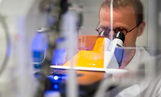
"First and foremost, our aim with this programme is to create a community of scientists studying the organisation of the cell nucleus in order to pool our analysis tools", noted Angela Taddei, a partner in this programme, director of the nuclear dynamics research unit (CNRS/Institut Curie). From this interdisciplinarity there should emerge innovative microscopes offering the possibility of monitoring molecules within a cell in real time and observing the dynamic of chromatin, the fibre composed of DNA and the molecules on which they coil. "The observation of these phenomena on the scale of a single cell should provide answers as to the levels of organisation of the genome, the way in which specific factors concentrate geographically without there being any compartmentalisation within the nucleus", said Maxime Dahan enthusiastically.
Apart from the development of microscopes, the scientists are researching probes which enable chromatin architecture or gene expression to be monitored at key moments in the life of the cells. "These are all tools which will enable us, for example, to monitor DNA repair mechanisms within the cells", explained Angela Taddei. This mechanism at the heart of her team’s studies eliminates errors arising in the DNA. Its failure leaves the door open to the emergence of cells which are potentially dangerous to the organism. Many other mechanisms should be revealed to the researchers in a new light thanks to these new means of observation of the cellular world.
Find out more
Coordinator :
- Maxime Dahan, head of the Curie's physical chemistry lab
Partners :
- Pr Edith Heard, head of the Curie's Genetics and Developmental Biology Lab,
- Angela Taddei, head of Curie's Nuclear Dynamics lab,
- Jean-Philippe Vert, leader of the Statistical Machine Learning and Modelling of Biological Systems' team,
- John Sedat of the Université de Californie (San Francisco, Etats-Unis) and Leonid Mirny from the MIT (Boston, Etats-Unis)
