• Home >
  • Teams >
  • Biology-inspired Physics at MesoScales

Presentation

Our research focuses primarily on the study of populations of interacting cells using physics concepts and techniques.

We conduct experimental projects addressing different aspects of communication between cells in different systems (including bacteria and mammalian cells). To achieve a good control in time and space on the local microenvironment of the cells, we extensively use the possibilities offered by microfabrication/micropatterning techniques. We also develop tools to quantitatively analyze our experimental data, which allows interpreting behaviors of populations on the basis of single cells’. The results obtained with this approach have led to several collaborations with theory groups and biology groups in France and abroad.


Collective chemotaxis of bacteria (A. Buguin)

Chemotactic bacteria E. coli swimming in suspension in a liquid medium consume the nutrients that are present in the solution and communicate between them via soluble factors. These properties give rise to complex behaviors (pattern formation, spontaneous fluctuations of concentration…). This system can be studied in microchannels, in which case, the bacteria form propagating waves when their concentration is sufficiently large. We measure quantitatively the propagation of these concentration pulses (figure 1) in various geometries of the channels and interpret these results with a kinetic model. Recent projects aim at perturbing these pulses by the application of an external field on the cells, or studying the interplay of mixtures of strains that behave differently in isolation.

Image Team Buguin

Figure 1: Concentration wave of E. coli bacteria in a microchannel and details of the individual trajectories

See our videos


Collective migration of epithelial cells (P. Silberzan)

When presented with a free edge, epithelial cells pertaining to a monolayer collectively migrate on the newly available substrate while maintaining cell-cell contacts (figure 2). For cells adhering to their substrate, protrusive activity controls this migration down to very small sizes. In contrast, for weak cell-substrate interactions, wounds close by a purse string mechanism via the peripheral actomyosin cable. Collective migration generally involves migration fingers driven by leader cells that develop at the free edge. These fingers composed of up to 100 cells, behave as mechanically coherent “super-cells” and reproduce at the multicellular scale the force dipole developed by migrating single cells. Collective cell migration is well described by an interacting particles model in which cells tend to adapt their motion to that of their neighbors. The same model also describes quantitatively the breathing oscillations observed in confined confluent epithelia at low cell density whereas, at high density, the movements of the cells gradually “jam” and tridimensional multicellular structures develop.

Image Team Buguin

Figure 2 : Top: Closure of a circular wound. Note the circumferential acto-myosin cable and the protrusions (stars). Bar=20µm. Bottom: Typical migration finger. Note the large leader cell and the actomysin cable at edges of the finger. Bar=50µm. (On both images: actin green, myosin red)

See also our videos.


Cellular Nematics (P. Silberzan)

Confluent spindle-shaped cells (such as fibroblasts or myoblasts) form monolayers presenting domains of common orientation separated by topological defects characteristic of nematic phases (Fig. 3). Plating fibroblasts on micro-patterned stripes yields large-scale, defect–free, perfectly aligned assemblies. In contrast, confining the cells on disks results in only two defects whose positions are characteristic of a system dominated by cell-substrate friction. Myoblasts plated on stripes develop an instability above a critical width, characterized by the onset of a chiral tilt of the cells relatively to the stripe’s direction together with a spontaneous shear flow of the cells at the borders. These results are consistent with active gel theories developed in the laboratory. We now address the importance of this contribution to cocultures of epithelial and nematic cells.

Image Team Buguin
Figure 3. Orientation of fibroblasts in a monolayer. Note the large nematic order and the presence of defects that prevent macroscopic alignment. Image size = 1560µm.

See our videos


Cell Competition (I. Bonnet)

Cell competition is a fundamental process by which interactions between two cell types direct the elimination of one of them. Up to now, experimental work has mostly addressed the behavior of transformed single cells within normal monolayers. We have recently started a project aiming at studying the behavior of an ensemble of cells transformed with the oncogene Src within a normal monolayer. For that purpose, we use an optogenetics-based set-up where an islet of cells of given shape and size can be transformed at will under the microscope by selectively exposing them to light at a specific wavelength. First results show a massive collective extrusion of the Src cells when surrounded by normal cells (Figure 4).

Image Team Buguin
Figure 4: Global collective extrusion of cells expressing the oncogene Src, embedded in a non-transformed monolayer. The local transformation has been performed with light.


Cell monolayers in topological complex environments (S. Coscoy)

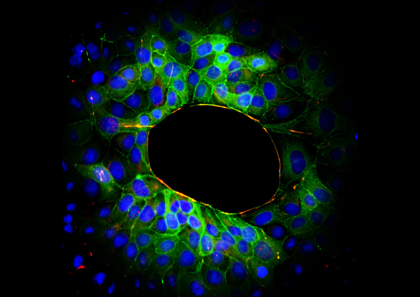
In physiological situations, epithelial cells in monolayers explore the third dimension by wrapping and folding this monolayer in space, to form for example tubules or cysts. We have recently investigated the impact of out-of-plane curvature on epithelial cells by plating them on glass wires of well-defined radii. The inverse geometry (growing the cells in tubes) is more relevant to study the mechanism of renal cyst formation that occurs in pathological situations. We have designed and developed a microfluidic set-up where cells are allowed to colonize cylindrical channels in hydrogels (Figure 5). This model system holds great promise to mimic the geometry and mechanical properties of kidney tubules.

Image Team Buguin
Figure 5: Kidney-derived cells lining the walls of 100 µm tubes managed in a collagen hydrogel.


More

Publications

2022Chiral Edge Current in Nematic Cell Monolayers

Physical Review X - 14/11/2022

See authors

2018Spontaneous shear flow in confined cellular nematics

Nature Physics - 01/07/2018

See authors

2017Collective cell migration: a physics perspective

Reports on Progress in Physics - 01/07/2017

See authors

Life of the team

Join us

Join our team!

There is currently no job offer.

See more job offers from the Research Center

Partners

See all teams