Team
Genetics of Tumor Suppression
Presentation
The p53 pathway is impaired in most, if not all, tumors. In half of human cancers the p53 gene is mutated, and in the other half the p53 protein can be inactivated by overexpression of one of its specific inhibitors, MDM2 or MDM4.
The p53 pathway is impaired in most, if not all, tumors. In half of human cancers the p53 gene is mutated, and in the other half the p53 protein can be inactivated by overexpression of one of its specific inhibitors, MDM2 or MDM4. A better understanding of the p53 pathway could lead to the development of new anti-tumor therapeutic strategies, applicable to many patients. Our group is developing mouse models to better understand the regulation and functions of p53.
We notably created the p53ΔP mutant mouse, expressing a p53 lacking its proline-rich domain, which proved to be very informative. The study of this mouse showed that MDM2 and MDM4 have distinct and complementary roles in the regulation of p53, and that the simultaneous inhibition of MDM2 and MDM4 is a strategy of choice for reactivating p53 in some cancers (Toledo & Wahl , Nat. Rev. Cancer 2007). Consistent with our results, ALRN-6924, a molecule that inhibits both MDM2 and MDM4, later showed its efficacy against some lymphomas (Ng et al., Nat. Commun. 2018).
We also created a mouse overexpressing the MDM4-S transcript, frequently overexpressed in human tumors. This model showed that MDM4-S is a marker rather than an important player in tumor progression. But more importantly, it indicated that the alternative splicing event leading to MDM4-S expression can lead to an activation of the p53 pathway (Bardot et al., Oncogene 2015). In agreement with our results, promoting this alternative splicing of MDM4 now appears as a promising strategy against melanoma (Dewaele et al., J. Clin. Invest. 2016).
These studies demonstrate the wealth of information acquired by studying the regulation of p53 in vivo, and the potential of these approaches for the development of new therapeutic strategies.
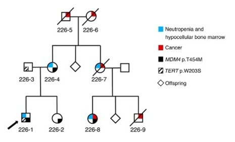
Our mouse models have also revealed unsuspected functions for p53. Indeed, we showed that a nonsense mutation leading to the loss of the p53 C-terminal domain leads to an increase in p53 activity, which causes bone marrow failure and pulmonary fibrosis. In humans, the combined observation of bone marrow failure and pulmonary fibrosis is characteristic of dyskeratosis congenita, a severe syndrome caused by telomere dysfunction. This led us to show that p53 is a major regulator of telomere biology, through its ability to repress the expression of several key genes, including DKC1 and RTEL1, two genes whose mutations can cause dyskeratosis congenita (Simeonova et al., Cell Rep. 2013 ; Figure 1). We next showed that a mutation leading to a partial inactivation of MDM4, and thus the overactivation of p53, can cause telomere dysfunction in humans (Toufektchan, Lejour, Durand et al. Sci. Adv. 2020).
These results led us to study how p53 represses the expression of genes important for telomere biology, and to identify other genes repressed by p53. We showed that p53 represses most of these genes indirectly, via the E2F4 repressor belonging to the DREAM complex. By the same mechanism, p53 represses the expression of several genes of the Fanconi DNA repair pathway (Jaber, Toufektchan et al., Nat. Commun. 2016), and genes important for centromere structure (Filipescu, Naughtin et al., Genes Dev. 2017). These results notably revealed FANCD2 and HJURP as markers of tumor progression, and suggested new therapeutic strategies.
Taken together, these results validate the medical relevance of our approach, which is based on the creation and analysis of mutant mouse models of the p53 pathway.