- Home >
- Institut Curie News >
- Embryo fragmentation under the microscope
In the very early days of our existence, cells from human embryos may fragment. This phenomenon is a common one and affects the embryo’s survival, particularly during procedures of assisted reproduction technology (ART). Cell fragmentation remained a mystery since mouse embryos, typically used to study human development, only rarely forms these fragments.

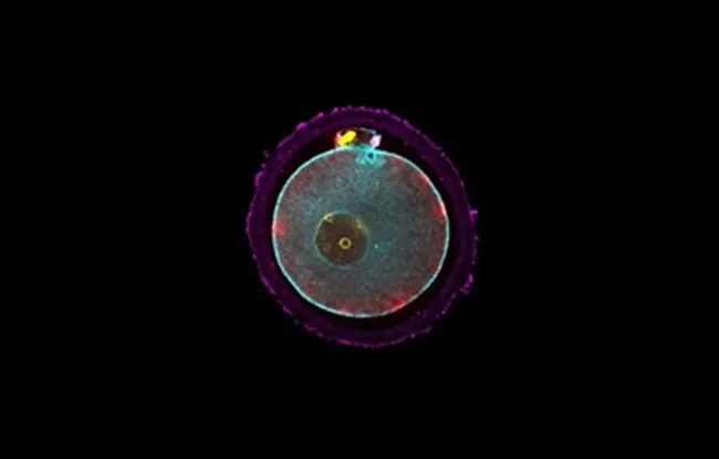
The team led by Dr. Jean-Léon Maître, CNRS research director and team leader at the Institut Curie in the Genetics and Developmental Biology unit (Institut Curie, CNRS, Inserm), made the serendipitous discovery that these fragments occur more frequently in the mouse after destabilization of the mitotic spindle. The mitotic spindle is the structure separating the chromosomes in cell division. Using a “light-sheet” microscope, they observed the formation of these fragments in three dimensions over time on embryos whose different cell parts had been fluorescently stained. These sequences were filmed and revealed the processes leading to cell fragmentation.
During cell division, if chromosomes are not properly attached to the mitotic spindle, they may get abnormally close to the surface. When chromosomes approach the cell surface, they trigger a contraction, very much akin to a muscular one, that pinches a piece of the cell and forms a fragment.
This sequence of events is reminiscent of another biological process, namely the formation of the polar body when the oocyte is formed. In order to form an oocyte containing sufficient resources for the embryo, the last division of the egg, known as “meiotic”, ejects half of the mother’s genome into a mini-cell, called the polar body. The signals coordinating formation of the polar body are well known: the chromosomes are taken close to the cell surface and trigger a cascade of biochemical reactions that leads to a contraction that pinches a small piece of the cell to form the polar body. However, these signals are assumed to be active only during this phase of “meiotic” division of the oocyte, and should not be present during “mitotic” divisions of the embryo.
By observing these biochemical “meiotic” reactions in the embryo under the microscope, scientists found that they are present during embryo fragmentation. Thus these signals persisting since the formation of the oocyte are the cause of the problems suffered by the embryo. These signals could be potential targets for improving ART, which are involved in over 20,000 births (~3%) a year in France.