- Home >
- News
Institut Curie News
Discover the latest news from Institut Curie.
Highlights

Search
Filter
Filter
Publication date
Fields
Search
240 result(s)
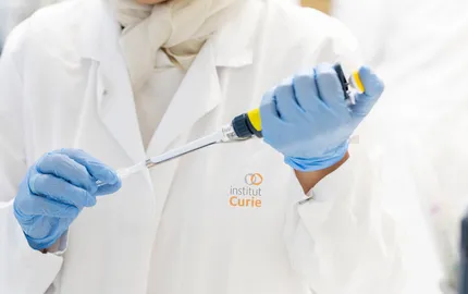
- Innovation
International women's rights day: Institut Curie commits to women's entrepreneurship
05/03/2026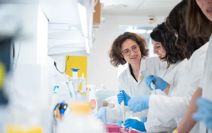
- Paediatric cancers
ERN PaedCan: Strengthening Cross-Border Care for Children and Adolescents with Retinoblastoma in Europe
17/02/2026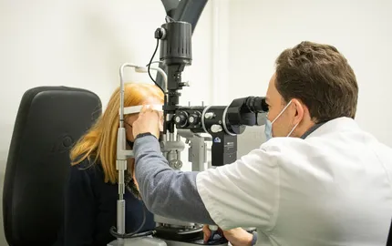
- Celebration
The Immunity and Cancer research unit (U932) celebrates its twentieth anniversary
12/12/2025
- Artificial Intelligence
SHAIPED: Institut Curie integrates a consortium of experts to create a "European space for AI in health”
08/12/2025
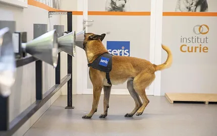
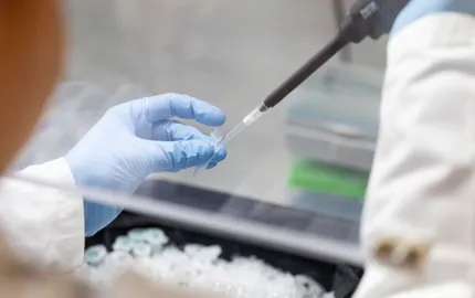
- Clinical research
From canine odorology to electronic noses: a new way to detect breast cancer
27/11/2025