- Home >
- Institut Curie News >
- Brain Metabolism and Lung Cancer: The Brain Provides New Keys for Determining Prognosis
In recent years, we have seen significant progress in the management of cancer patients, in particular due to the precision with which tumors are characterized, allowing treatments to be adapted to each specific case. As part of a system-wide medical project, which tends to take into account the patient's situation as a whole, a retrospective study conducted at Institut Curie showed that in patients with lung cancer, cerebral hypometabolism is associated with a poor prognosis. Published in The Journal of Nuclear Medicine in April 2026, this study opens the way for promising research avenues and the development of new prognostic tools.
Lung cancer is the 2nd most common cancer in men and the 3rd in women, with nearly 53,000 new cases per year in France1. Throughout the course of patient care, medical images, including those obtained by PET Scan2, make it possible to monitor the course of the disease.
"In these images, only a limited number of parameters measured on the tumor lesions are taken into account by the doctors," explains Dr. Irène Buvat, CNRS research director and director of the new Imaging, Innovative Radiotherapy and Systems Medicine unit (IRIS - Institut Curie / CNRS UMR9029 / Inserm U1353 / Versailles Saint-Quentin University). "However, the patient's prognosis cannot be reduced to just the tumor lesions... That is why we examined the surrounding organs and tissues to assess whether their functioning could also be impaired in the presence of cancer and to provide updated information about the patient's condition. Understanding these links could enable us to propose additional criteria for monitoring disease progression—or the lack thereof—based on tests which were already performed during the course of patient care."
A retrospective study of 380 patients identifies new avenues for biomarker research
A study carried out by Dr. Julie Auriac as part of her thesis and directed by Dr. Irène Buvat and Dr. Fanny Orlhac, Inserm Research Officer and Head of the Integrative Radiomics for Precision Medicine team within the IRIS unit, has been published in the Journal of Nuclear Medicine on April 9, 2026. Clinical and imaging data from 380 patients with non-small cell lung cancer (about 85% of lung cancers 3) treated at the Curie-Montsouris Chest Center between 2010 and 2023 were retrospectively analyzed to identify correlations with patient survival.
"This retrospective study allowed us to explore new avenues. This would have been impossible a few years ago. It is thanks to the AI algorithms that help us automatically annotate the images and extract the parameters that we were able to discover this unexpected link between brain metabolism and prognosis, " adds Dr. Fanny Orlhac.
Brain metabolism: a new prognostic tool?
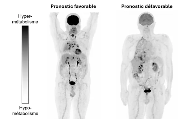
By exploring the clinical, biological, and imaging data of patients, the researchers were challenged by one parameter in particular—brain metabolism. Just like tumors, the brain consumes a lot of glucose. On the PET scan, it shows strong uptake of the radiotracer. By quantifying this, the researchers observed that a lower uptake was associated with a higher mortality risk.
"In other words, when brain metabolism is reduced, survival is also reduced. But the reasons for this decrease remain unknown. We know that brain metabolism can be affected by many factors, such as age or inflammation. We have ruled out these hypotheses, and we believe that it is likely a marker of the general functional state of the patient," suggests Dr. Irène Buvat.
"Indeed, the patient's physical, psychological, or nutritional status, as well as the treatments administered, can influence cerebral metabolism. However, we did not have systematic access to this type of information, as the study was retrospective. These are the avenues that we will explore later, prospectively collecting information that could allow us to explain this variation in brain metabolism in patients with poor prognosis," explains Dr. Fanny Orlhac.
Towards systems-based medical research
A new vision of the disease, and the impact it has on the body, has been emerging for the past few years: the opinion that the body is affected by disease in its entirety. It is a paradigm shift that tends towards research that is no longer focusing only on the diseased organ, but on the whole person 4. "Medicine and research are fragmented today: each organ is studied separately from the others and specialists in different organs do not interact frequently. Today we propose a profound paradigm shift, a reorganization of culture and research methodology in order to understand how a disease can affect the whole body, " specifies Dr. Irène Buvat. "In practice, this requires interdisciplinary research, a pillar of the teams' work at Institut Curie. Our new IRIS unit employs researchers, doctors, and engineers from different backgrounds and different disciplines, and this proximity provides us real impetus in our research projects," concludes Dr. Fanny Orlhac.
1 Source
2 PET scan: Positron emission tomography (PET) imaging makes it possible to visualize the functioning of organs and tissues. In oncology, the most frequently used radiotracer provides information on the glucose consumption of cells and thus makes it possible to identify tumor lesions, evaluate their extent, as well as assess the response to treatments
3 Source
4 Buvat et al, 2026 Cross-disciplinary methodologies for whole-person research– insights from EMPOWER2024