- Home >
- Institut Curie News >
- Cell division and cell geometry
Round, elongated in a pear or hexagon shape, the shape of cells is essential to life! Every day millions of cells divide in our bodies. For each division to succeed, the cell must alter the geometry of its cytoskeleton to the nearest micrometer. It ensures the precise segregation of the genomes of the daughter cells and prevents tumors from occurring.
However, the way in which these geometry changes take place in the epithelia was poorly understood. To answer this question, we combined the power of genetics of the fruit fly with time-lapse imaging
Explains Yohanns Bellaïche, head of the Polarity, Division and Morphogenesis team and Deputy Director of the Genetics and Developmental Biology unit (CNRS UMR 3215/Inserm U934/Sorbonne University) at Institut Curie.
The work was conducted at Institut Curie’s Research Center in collaboration with the research team of Eurico Morais-de-Sá from the Instituto de Investigação e Inovação em Saúde da Universidade do Porto (I3S) in Portugal.
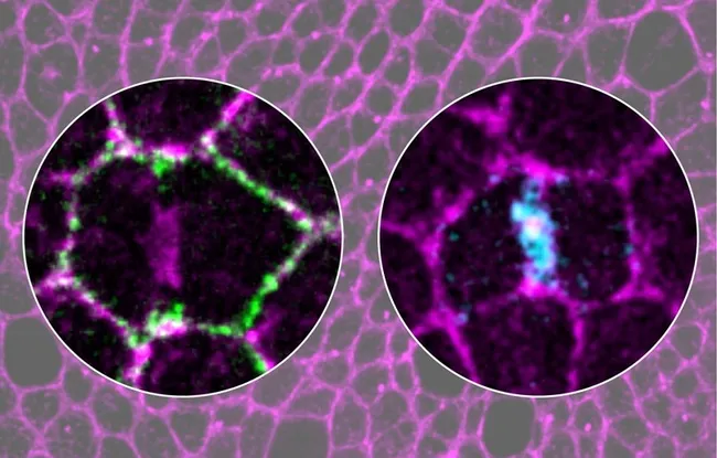
By recording image by image the dynamics of a large number of proteins, during cell divisions in the epithelia, we revealed a complex choreography of proteins, the RhoGTPases
Continues Y. Bellaïche.
These proteins remodel the cell’s actomyosin cytoskeleton and thus control cell geometry. This molecular choreography gives rise to a ballet where each dividing cell coordinates its geometry changes with the cells surrounding it. The result is that the two daughter cells are in the right place and well attached to their neighbors.
Using high-resolution imaging, we revealed the mechanisms that activate these RhoGTPases, which help epithelial cells to divide correctly
The team leader continues.
But the researchers are very much aware that the slightest misstep in this ballet can lead to a tumor site.
Systematic analysis of Drosophila RhoGEF/GAP localizations uncovers regulators of mechanosensing and junction formation during epithelial cell division
Florencia di Pietro, Mariana Osswald, José M De las Heras, Ines Cristo, Jesus Lopez- Gay, Zhimin Wang, Stéphane Pelletier, Isabelle Gaugué, Adrien Leroy, Charlotte Martin, Eurico Morais-De-Sá, Yohanns Bellaïche. Current Biology, 13 février 2023 https://www.cell.com/current-biology/fulltext/S0960-9822(23)00062-3
Support from the ERC
This team’s project received support from the European Research Council (ERC) to develop an artificial intelligence IT tool that learns to recognize, using microscope images, when a biological phenomenon is about to occur, and then adapts the way the picture is taken, by zooming in, or taking a succession of images closer together in time.