- Home >
- Institut Curie News >
- Shadocks and embryos, same fight
The lumen of the embryo is formed from hundreds of mini-lumens
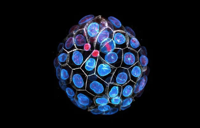
Brain, heart, liver, kidneys, most of the organs in our body contain cavities filled with fluid. These cavities called lumens are essential for the proper functioning of our organs. During the very first days of our development, when it consists of only 32 cells, the embryo forms its first lumen. This lumen swells inside the embryo and its position determines the place through which the embryo will attach to the maternal uterus to implant and continue its development.
In 2019, a study led by the Dr. Jean-Léon Maître, head of the Mechanics of mammalian development team (CNRS UMR3215 / Inserm U934 / Sorbonne Université), showed that the lumen is formed by hydraulic fracturing: the pressure of the fluid breaks the contacts between the cells. But this fracturing does not form a single lumen but hundreds of mini-lumens, or pockets, which gradually exchange their contents and empty into each other until a single dominant lumen accumulates all the fluid. Thus, these flows between pockets are crucial for positioning the lumen and determining the implantation point. But how do the cells control the fluid movements within the embryo?
Thousands of pumps, the blebs reversed, to ensure the formation of the lumen
In an article published in the journal Nature Cell Biology, the same scientists discovered structures called inverted blebs that pump fluid from one pocket to the other. These inverted blebs swell with fluid in 10 seconds to reach a few micrometers in diameter before contracting in 30 seconds to repel the fluid. To go from a network of pockets to a single large lumen in a few hours, thousands and thousands of inverted blebs appear and disappear within each embryo.
By manipulating the ability of embryos to accumulate fluid or to constrain the fluid in the pockets, scientists have shown that inverted blebs are formed thanks to the pressure of the fluid which pushes like a balloon towards the inside of the cells. These rapidly recruit proteins from the actomyosin cytoskeleton (the same ones that allow our muscles to contract) on the surface of the inverted blebs and push the fluid back to other pockets. Thus, the inverted blebs pump and pump and pump again the fluid within the embryo, thus circulating it. But this back and forth of the fluid does not seem very productive.
The scientists then identified that some pockets, those surrounded by more than two cells, did not form inverted blebs with as high a frequency as the other pockets containing only two cells. In fact, the pockets surrounded by more than two cells would not pump efficiently and would act like a siphon. To support this hypothesis, the scientists manipulated the topology of the embryos in such a way as to eliminate the pockets surrounded by more than two cells. These structures without a siphon form a lot of inverted blebs but, as they pump the fluid from one to the other in a futile cycle, they only painfully manage to form a lumen. Thus, these structures without a siphon are like the Shadocks, one of the adages of which is "It is better to pump and nothing happens rather than not to pump and risk something worse happening”.
This study therefore highlights inverted blebs as the cellular mechanism allowing embryos to form a single lumen whose position determines its future development. She also reveals that to go beyond the first days of the development of the mammalian embryo it is essential to follow this other Shadockian maxim: "I pump so I am".
Schliffka, M.F., Dumortier, J.G., Pelzer, D.et al. Reverse blebs operate as hydraulic pumps during mouse blastocyst training. Nat Cell Biol(2024). https://doi.org/10.1038/s41556-024-01501-z

Figure: Schematic diagram of lumen formation in the mouse embryo. Fluid (blue) accumulates between the cells inside the embryo. The fluid-filled pouches exchange their contents until only one pouch remains, forming the lumen that determines the embryo's axis of symmetry and point of implantation. To help the pouches exchange fluid, structures called inverted blebs (green) pump fluid from one pouch to the other.
Research News
Discover all our news
Celebration
The Immunity and Cancer research unit (U932) celebrates its twentieth anniversary
12/12/2025
Artificial Intelligence
08/12/2025


