- Home >
- Institut Curie News >
- Under Pressure: Toward a Better Understanding of Caveolae
Caveolae are small pouches found on the surface of cells. Long thought to be involved solely in intercellular communication, caveolae also enable adaptation to mechanical stress. Like an accordion, caveolae can unfold, allowing the cell to respond dynamically to surrounding physical forces. At the intersection of physics and biology, a new study led by Drs. Christophe Lamaze and Cédric Blouin sheds light on the cascade of events triggered by the unfolding of caveolae. These findings were published in Nature Cell Biology on June 1, 2026.
The body’s cells are subjected to various types of mechanical forces. This is the case, for example, with tumor tissues, which grow and press against surrounding cells, abnormally increasing their membrane tension. In response, cells are able to normalize their plasma membrane tension through specialized membrane structures called caveolae.
Caverns and the regulation of membrane tension
“Caveolae were discovered in the 1950s, but it is only recently that we have come to understand their true roles. And we owe this to the physicists at Institut Curie! Our collaboration with theoretical physicist Pierre Sens1 , one of the authors of this new study, began following discussions during the Scientific and Medical Day,” says Dr. Christophe Lamaze, Inserm research director and head of the Membrane Mechanics and Dynamics of Intracellular Signaling team in the Chemical Biology of Cancer unit (Inserm U1339 / CNRS UMR3666).
The seminal study was published in 2011, laying the groundwork for a new understanding of caveolae. These structures detect changes in membrane tension and, by unfolding, enable the membrane to withstand stress and prevent it from rupturing. However, much remains to be understood about the mechanisms involved in this mechanical sensitivity function.
Intracellular signaling
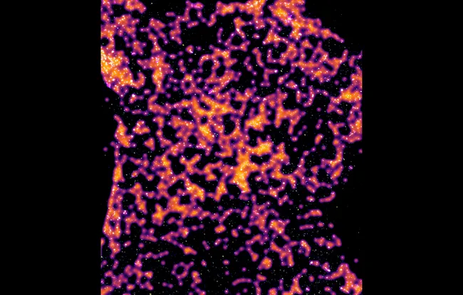
The team therefore sought to understand what happens inside the cell once the caveolae unfold. In collaboration with Gregory Giannone’s team in Bordeaux and Robert Nabi’s team at the University of Vancouver, they used super-resolution microscopy techniques and artificial intelligence algorithms to analyze the signals. They were thus able to show that the disk-like components of caveolae diffuse and can interact directly with other proteins in the cytoplasm, regulating their activity.
“We show that caveolae components, released by mechanical stress, inhibit a cellular signaling pathway called JAK/STAT, a pathway central to tumor and immune processes,” explains Dr. Cédric Blouin, an Inserm research fellow in Christophe Lamaze’s team.
“We are discovering more and more links between dysfunctions in caveolae-mediated mechanical sensitivity and the development of diseases. This is the case, for example, with cancer: overexpression of CAV1, which encodes a protein that forms caveolae, promotes metastasis and resistance to treatment. A better understanding of caveolae and the consequences of their dysfunction in pathophysiology could lead to the development of new avenues for research and treatment,” concludes Christophe Lamaze.
[1] Research director at the CNRS and Physical Approach of Biological Problems team leader in the Physics of Cells and Cancer unit (CNRS UMR168 / Sorbonne Université)
Image Caption: STORM images with caveolin-1 (fire LUT) and JAK1 (blue/yellow LUT). The « influence zone » of caveolin-1 is indicated with blurry areas where JAK1 should not be active.