Presentation

Chromatin organization in the nucleus provides a large repertoire of information in addition to that encoded genetically. Understanding how this information is established and possibly inherited through cell division is a challenge for the field. A key question is how histones, the major protein components of chromatin, as particular variants or post-translationally modified forms, can mark functional regions of the genome.
Our team is interested in understanding how chromatin organization is established, propagated, maintained, and changed during development and in response to environmental cues. Errors in these processes can lead to mis-regulation of genome functions and pathological outcomes, such as cancer.
Our general objective has been to dissect the mechanisms of chromatin assembly, from the basic structural unit, the nucleosome, up to higher-order structures in the nucleus (Fig. 1). We have characterized key chaperones involved in nucleosome assembly and defined the dynamics of new histone incorporation in chromatin. Our findings have shed light on the fundamental issues of the dynamics, fate, and inheritance of histones, with their specific marks typical of particular chromatin domains.
Our working hypothesis is that histone chaperones function in an ‘assembly line’ with specificity for individual histone variants to mark defined regions of the genome. Remarkably, we have found that misregulation of specific histone chaperones is a common feature of aggressive breast cancers.
Our plan is to analyze the regulatory pathways that target histone chaperones and variants to control the assembly line and its connecting network.
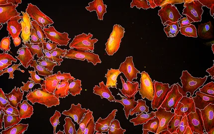
Our specific approach to understanding all the in vivo functions of chromatin complexes is based on tools and model systems (e.g. Xenopus, mouse) that combine biochemistry, cell biology, and developmental biology (Fig. 2). We examine specific nuclear domains: non-coding centromeric heterochromatic regions, which are of major importance for chromosome segregation.
Together these studies should ultimately help in the development of medical applications of relevance for cancer.


