Presentation

Team organization
SAIRPICO Project-Team, Centre Inria de l’Université de Rennes, Campus Universitaire de Beaulieu, 35042 Rennes Cedex, France and Institut Curie Research Center, Cellular and Chemical Biology unit (CNRS UMR3666 / INSERM U1143), 26 Rue d'Ulm, 75005 Paris.
Based on more than 10-year shared experience and the foreseen benefice of grouping people from cell biology, microscopy, image processing and analysis, and statistical learning, the SERPICO Project-Team (2017-2023) and the SAIRPICO Project-Team (since April 1st, 2023), located in Rennes and Paris, involving Inria (Centre of University of Rennes), Institut Curie Research Center (CNRS UMR3666 / INSERM U1143), PSL Research University, develop statistical and computational methods for cell imaging, and biomedical research. Since 2023, the new interdisciplinary team, strongly supported by the aforementioned institutions, is composed of 6 researchers (artificial intelligence, chemo-biology), 6 PhD students (statistical learning and image analysis), 2 postdocs (statistical learning, deep learning), and 2 engineers (computer science, chemo-biology).
See the full list of team members
Scientific context and objectives
All fluorescence microscopy systems record fluorescent signals emitted by tagged molecules. Fluorescence information necessarily includes intensity (biomolecule density), wavelength (absorption and emission spectrum), time (fluorescence decay lifetime) and polarization (which arises from the dipole orientation). The next generation technology will be able to better reveal the structure and function of biomolecules in cells, and to decipher complex biological mechanisms. Among the recent progress, let us mention polarized microscopy that has the potential to probe the dipole orientation of fluorophores linked to proteins or lipids of interest and thereby, to provide another layer of spatial and environmental information at the nanoscale. As the corresponding image data are 3D+time multi-valued signals that could potentially depict several fluorescently tagged molecular species, the analysis of fluorescent microscopy images remains a challenge in signal-image processing and machine learning, in order for this technology to be adopted more widely in biological studies. These point to under explored areas of research in image processing and machine learning, to fully exploit the potential of cutting-edge microscopy in biological studies, our global objective is to develop the next generation of multi-valued information processing techniques. This objective will be achieved by integrating theoretical concepts in signal-image processing, innovative developments in optical engineering and chemistry, and the stringent requirements of cell imaging. The new generation algorithms based on statistical and machine learning will serve to characterize the dynamics of biomolecules and to decipher the molecular transport pathways or the motion and deformation of cells, which is of considerable of interest in fundamental cell biology and for precision medicine.
Research axes
Four complementary Research Axes will be investigated with scientists who develop chemical methods to infer and quantify interactions between membrane proteins.
Axis 1- Modeling and reconstruction of multi-valued images
Development of cutting-edge computational strategies and mathematical frameworks for reconstructing multi-valued images. Structure-based sparse representations of multi-value images will be established from the analysis of the spatiotemporal correlations and the inherent redundancy of data in multiple images. We will investigate statistical nonparametric methods and aggregation techniques, variational Bayesian methods, including shape-based models, as well as machine learning strategies to solve the underlying inverse problems.
Axis 2- Methods for spatial high-resolution quantification of molecular mobility and interact tions.
Characterization of molecular mobility at the nanoscale from multi-valued images. We intend to fully exploit the rich contents of microscopy images in order to build single-molecule (e.g., endocytic ligands) and biomolecule (e.g., cytosolic machinery, metabolic sensors) tracking algorithms, derive robust estimators of molecular mobility, and quantify spatially-variable interactions between molecular species and cytoskeleton. The resulting algorithms will be used to produce spatial high-resolution maps of molecular mobility given stochastic motion models and sparse representations.
Axis 3- Spatiotemporal modeling of 3D shapes, motions and deformations.
Development of shape models and descriptors to capture 3D motion and deformation of macromolecular complexes (cryo-electron tomography (cryo-ET), single particle analysis (SPA)) in one hand, and in the other hand, intracellular components and tumor cells, at the scale of a single cell and tissue-scale. We intend to represent 3D shapes by parametric surfaces controlled by key points and to segment and track structures in 3D microscopy. The main originality will be to exploit leverage annotations and/or high-level priors to derive features for classifying molecular conformations in cryo-ET, and phenotypes induced by drugs (single cell), or controlled hypoxia conditions (tissue-scale) in 3D+time fluorescence microscopy.
Axis 4- Analysis of case-studies in cell biology and cancer research.
Demonstration that the methods and algorithms related to the three previous methodological axes allow one to perform image reconstruction for several 3D instruments (TIRFM, Lattice Light Sheet Microscopy, Multi-Focus Microscopy, cryo-ET), and accurately quantify the shape and motion of cell components and biomolecules that interact with membranes and the cytoskeleton. The resulting images and features will be helpful to better decipher the intracellular dynamics of trafficking and signaling events in living cells, especially membrane mechanics at the cell surface, endocytosis, as well as signal transduction to the nucleus. The methods will be developed for investigation in cellular and chemical biology, and extended further to perform analysis at the tissue-scale.
Biosketch
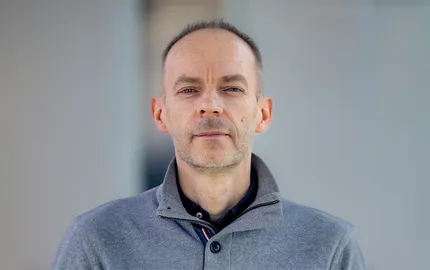
Charles Kervrann is Inria Research Director (DR1) and is heading the Sairpico Project-Team (Inria Rennes, Institut Curie, CNRS UMR3666, Inserm U1143, PSL University) jointly located in Rennes and Paris since April 1st, 2023. Previously, he was head of Serpico Project-Team (Inria, Institut Curie CNRS UMR144, PSL University) (2018-2023). He received his PhD and HdR in Signal Processing and Telecommunications from the University of Rennes 1 in 1995 and 2010, respectively. He was appointed INRA CR2 (Applied Mathematics and Informatics, Jouy-en-Josas) in 1997 and moved (on Inria secondment) to Inria Rennes in 2003. He was recruited as Inria Research Director (DR2) in 2010. His research interests are in the field of image processing and machine learning, inverse problems in biological imaging. He has supervised 19 PhD students, 7 postdocs and 6 engineers. He was Associate Editor of “IEEE Signal Processing Letters” (2015-2019), member of the IEEE BISP “Biomedical Image and Signal Processing” committee (2010-2018), and member of executive board of the CNRS-GdR ImaBio (2010-2020). He is currently Associate Editor of “IEEE Trans. Image Processing” and “Biological Imaging” journals and co-head (with Institut Pasteur) of the “BioImage Informatics” node within ANR PIA France-BioImaging since 2012. He was Co-General Chair and Organizer of the “Quantitative BioImaging” (QBI’2019) conference in 2019. He has coordinated several projects with academic teams: ANR PRME POLARISCOPIA (2022-2026); ANR PRC DALLISH (2016-2020, with Institut Curie and INRA); Inria DEFI NAVISCOPE (2018-2022, with 9 research teams); CATLAS (2016-2021, with Max Planck Institute Martinsried, Germany); CytoDI Associate-Team (2016-2018, with UTSW Medical School Dallas, USA). He has participated to additional projects supported by ANR or institutions in collaboration with Institut Curie (since 2005), IGDR Rennes (since 2005), INRAe MaIAGE and MICALIS Units (since 2017), Hôpital Saint-Louis Paris (2015-2016), IINS Bordeaux (since 2019), IRSN (since 2020), Institut of Neuroscience Grenoble (since 2021), Institut Gustave-Roussy (since 2022), IGBMC Strasbourg (since 2021), and LMJL University of Nantes (since 2016). At the international level, he has collaborated with University of Cambridge (UK, since 2019), CSIC Madrid (Spain, since 2020), Oviedo University (Spain, since 2022), Helmoltz Pioneer Campus (Neuherberg Germany, 2020-2021), MacMaster University (Canada, since 2021), Harvard Medical School (2013), University of California San Francisco (since 2010). He has collaborated with companies: Airbus Defense and Space (2020-2024), Myriade (2021), GATACA Systems (2016-2023), Nobleo/UVISIO (The Netherlands, 2020), OBSYS (2016-2018), INNOPSYS (2013-2017). He has published 60 publications in international journals, and 80 publications in peer reviewed conferences.


