

- Home >
- Institut Curie News >
- A new medium-field PET scanner at Institut Curie
Faster diagnosis, improved patient comfort... Institut Curie recently acquired a new, state-of-the-art medium-field PET scanner—a world first* from Siemens Healthineers. Following a meticulously planned installation, this innovative device has been treating patients since May. Read on to learn from Dr. Romain-David Seban how this exceptional installation unfolded and the benefits it will bring to the institute’s patients.
PET (positron emission tomography) imaging combined with CT (computed tomography) plays a vital role in the care of cancer patients, from initial diagnosis through follow-up during treatment. This technology makes it possible to visualize metabolic or molecular activity—particularly that of tumors—and thus helps doctors assess the progression of the disease and the effectiveness of treatments.
An extraordinary installation
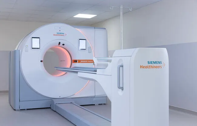
Committed to continuing to offer its patients the most advanced technologies, Institut Curie recently acquired a new mid-field PET-CT scanner (Biograph Trinion.X* model1 ). This is the very first machine of this model installed worldwide by Siemens Healthineers. This equipment is now available at the Saint-Cloud site.
“Installing a machine like this is a real technical challenge that requires meticulous preparation and coordination among many professionals. As a first step, work was carried out to adapt the facilities and prepare for the arrival of this equipment,” explains Dr. Romain-David Seban, head of the Nuclear Medicine Department in Saint-Cloud.
The machine was delivered in several parts before being assembled on site. “We then implemented a phased rollout with a limited number of patients. This step allowed medical radiology technologists (MERM) to familiarize themselves with the machine’s new features, while ensuring optimal and safe patient care,” explains Dr. Romain-David Seban.
Cutting-edge technology for the benefit of patients
This new PET-CT scanner features a wider axial field of view: “In practical terms, the machine can acquire images of a larger area of the body in a single scan. This reduces the duration of exams, improves patient comfort, and optimizes department operations,” explains Dr. Romain-David Seban.
Thanks to its increased sensitivity and state-of-the-art detectors, this PET-CT scanner also produces more precise images and provides better visualization of certain small lesions. Another significant advantage is that image quality can be maintained while reducing the amount of radioactive tracer injected into certain patients. “Beyond its clinical use, this machine will also open up new avenues for research conducted at Institut Curie. I am thinking in particular of the research being done in the Imaging, Radiotherapy, and Systems Medicine Unit , led by Dr. Irène Buvat, with which I am affiliated for my research activities. Thanks to these new imaging capabilities, we will be able to better leverage the information contained in medical images to improve patient follow-up and, ultimately, contribute to increasingly personalized care,” concludes Dr. Romain-David Seban.
Check out the video presentation of this new PET scanner
*Biograph Trinion.X EP9 paired with a 64-slice CT scanner
