- Home >
- Institut Curie News >
- What Physics Teaches Us About the Role of Cellular Organization Defects in the Birth of a Three-Dimensional Tissue
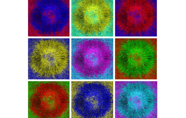
How can a single layer of cells evolve into an organized mass? And can we predict where this transition will take place? A single defect in the collective alignment of cells is enough to spontaneously trigger the formation of such an organized 3D structure several layers thick. This is what the work of the PhysicoBiology at Mesoscales team (CNRS UMR168 / Sorbonne University) led by Dr. Pascal Silberzan, CNRS Research Director of Institut Curie reveals. A discovery published in Nature Physics that could partly explain the development of tumors?
For Dr. Pascal Silberzan1, the proximity between physicists and biologists within Institut Curie is an opportunity. "Physicists like me are sensitive to biological issues while having a different perspective than our colleagues. In the team, we study the collective behavior of cell populations, rather than the functioning of an isolated cell. Imagine fish swimming together. We are interested in the movement of the school, less so in the mechanisms used in the swimming of each fish."
To study this collective cell behavior, the team, funded by the League Against Cancer, is working closely with theoretical physicists from the Physical Approaches of Biological Problems team, led by Dr. Pierre Sens within the same research unit. She works in particular with Dr. Carlès Blanch-Mercader and Dr. Jacques Prost, whose models have made it possible to interpret the observations.
Cells that behave like a liquid crystal
In practical terms, the team cultivates myoblast monolayers2. These elongated cells constantly exert mechanical forces on one another and on their substrate. Physicists speak of "active matter" and model these monolayers as a gel with these mechanical characteristics. In addition, these cells spontaneously align with each other, in the manner of liquid crystal molecules3.
But this alignment cannot propagate indefinitely, and the monolayers locally trap organized defects. "These have long been underestimated in biology, as it is tempting to prioritize studying homogeneous regions," explains Pascal Silberzan. Now, physicists who are well-acquainted with these intrinsic defects in liquid crystals know that they are potentially the seat of interesting properties. It is also known that they are the seat of important collective forces and cellular expulsions outside the monolayer, as recent work has shown4."
A custom-made vortex
To understand what is happening at the level of a defect, the team chose to impose it artificially. Using microfabrication techniques from microelectronics, researchers etch concentric tracks onto a surface, spaced a few microns apart, creating a pattern of concentric circles (a "vortex"). And thanks to confocal microscopy and video microscopy from the CurieCoreTech Cell and Tissue Imaging (PICT) platform at Institut Curie, they observe that the cells, guided by this relief, orient themselves along the tracks and follow them faithfully. But as the density increases, a massive convergent flow overlays this contact guidance, drawing the cells from the periphery toward the center of the vortex, over several millimeters. “On these circular rails, the curve is sharp in the center and gentler on the outside. The cells are therefore subjected to a curvature gradient, and this gradient imposes a force perpendicular to the rails that pushes the cells toward the center of the vortex," explains Carles Blanch-Mercader, who contributed to the study at Institut Curie.
After 12 hours, cells begin to escape from the plane at the center of the vortex, forming a second layer, then a third. And each new layer in turn becomes the seat of a convergent flow that feeds the next one. In a few days, a cellular mound about ten cell layers high is thus formed.
A perfectly ordered structure
The discovery of this mechanism, where each layer develops by cell transfer from the layer below, is itself a first. But that's not all: by following the evolution of these clusters, the team observes that the stacking spontaneously reorganizes. The first layer remains vortex-oriented, the intermediate layers spontaneously take on the star-like shape of an "aster," while the top curls into a spiral. "It's a very reproducible structure that shows the tissue has the capacity to modulate its defects," insists Dr. Hsiang-Ying Chen, a postdoctoral researcher in Pascal Silberzan's team at the time of the study, and first author of the publication. Only the core of the structure forms an unordered line between the initial defect and the top of the mound.
Possible avenues for cancer research
Falling under basic research, this work does not reproduce cancer mechanisms, but "it could shed light, by analogy, on the transition from a two-dimensional tissue to three-dimensional growth, a central phenomenon of tumorigenesis," notes Pascal Silberzan cautiously. If this hypothesis is confirmed, it would open up an original therapeutic lead. Instead of targeting only tumor cells, why not aim to disrupt their ability to organize collectively?
[1] CNRS Research Director and Head of the PhysicoBiology team at Mesoscales (CNRS UMR168 / Sorbonne University) at Institut Curie.
[2] Myoblasts are stem cells responsible for the formation of skeletal muscles in the embryo.
[3] A liquid crystal is a state of matter that combines the properties of an ordinary liquid with those of a crystalline solid. Here, the cells are aligned with each other but arranged randomly in the monolayer.
[4] Sarkar T., Yashunsky V., Brezin L., Blanch Mercader C., Aryaksama T., Lacroix M., Risler T., Joanny J.-F., Silberzan P.: Crisscross multilayering of cell sheets, PNAS Nexus, 2, (2023), pgad034
Source
Reference: Chen H.-Y., Blanch-Mercader C., Giuglaris C., Prost J., Silberzan P., Morphogenesis of stratified cell mound at a vortex defect, preprint deposited on HAL on July 7, 2026, https://rdcu.be/onrPwVm2a9ja.
Nature Physics in press