- Home >
- Institut Curie News >
- Cryomicroscopy: the Research Center thinks big
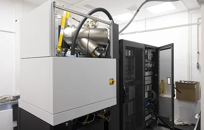
After several months of coordination by Dr. Daniel Lévy, Scientific Director of the Cell and Tissue Imaging Imaging Platform (PICT) in Paris, and the members of his Molecular Microscopy of Membranes team (CNRS UMR168/Sorbonne University), in collaboration with the technical departments and the information systems department, Institut Curie’s Research Center welcomed the brand new Glacios Cryo-TEM™ by Thermo Fischer in February.
Installed in the basement of the Pavillon Curie in the Physical Chemistry Curie unit (CNRS UMR168/Sorbonne University) headed by Dr. Pascal Hersen, this powerful cryo-electron microscope joins the state-of-the-art equipment dedicated to live imaging already in the PICT platform. Its cost of over 2 million euros was funded by Institut Curie’s Research Center, the Ile-de-France region, Institut Pasteur, CNRS and FranceBioImaging and in partnership with Institut Jacques Monod. An amount of 300,000 euros was also invested in renovations, thanks to the generosity of donors, to create room adapted to control vibrations and ambient magnetic fields.
Maintaining a significant level of investment is a recognition of excellence for the Research Center. The acquisition of this type of state-of-the-art equipment clearly boosts the status of our laboratories.
Explains Prof. Alain Puisieux, its director.
A unique microscope enabling new research approaches
This cryo-electron microscope enables observation at atomic resolutions (10-10 m) of the proteins involved in the main cellular functions, and which have a biomedical interest. Using cryo-microscope images, it is possible to reconstruct models of their structure and their environment. This helps us better understand how they function and the reasons for their deregulation, and can help with the design of drugs, particularly for cancer treatment.
We will be able to observe isolated proteins, presenting mutations, or combined with a drug, or the proteins present in the vesicles secreted by the immune or tumor cells. We will also be able to observe protein assembly responsible for remodeling of cell membranes during migration, cell division or intracellular traffic. It will then be possible to place all of this in an even broader context, in a cell, in order to understand the fine-tuned mechanisms underlying the main cellular functions and their pathological disruptions.
Explains Dr. Daniel Lévy
In allowing the structure of a protein and its close environment to be observed, this cryo-electron microscope thus represents the missing link between structuralists, biologists and physicists at Institut Curie.
Several research projects are already planned. For example, Dr. Aurélie Bertin, CNRS Research Director in Dr. Daniel Lévy’s team, is looking at the filaments formed by septins, cytoskeleton proteins, on the cell membranes, which leads to parent cell-daughter cell division. The cryo-electron microscope will provide a 3D vision of each of the filaments and deformations of membranes at short and long distance, and will enable her to model certain steps in cell division.
The team of Dr. Anne Houdusse-Juillé (CNRS UMR144/Sorbonne University) will determine the structures of molecular motors to understand the principles underlying the conversion of chemical energy into mechanical movement by these nanomotors and the mutations responsible for diseases.
The team of Dr. Clotilde Thery (Inserm U932/Université Paris Cité), is working on the extracellular vesicles secreted by the cells. Cryo-electron microscopy will provide a new vision with unprecedented resolution and ultimately help develop their therapeutic potential.
Lastly, the team of Dr. Graça Raposo (CNRS UMR144/Sorbonne University) will be able to correlate the cell images in electron microscopy with fluorescent images to have a comprehensive vision of intracellular traffic, at different scales.
Other research projects are also already in progress in the teams of Dr. Stéphanie Descroix, Dr. Ludger Johannes, Dr. Christophe Lamaze, and Dr. Patricia Bassereau.
This is a major and rare investment, but a necessary one for developing new innovative projects and sustaining the excellence of our research
Concludes Dr. Pascal Hersen, director of UMR168.